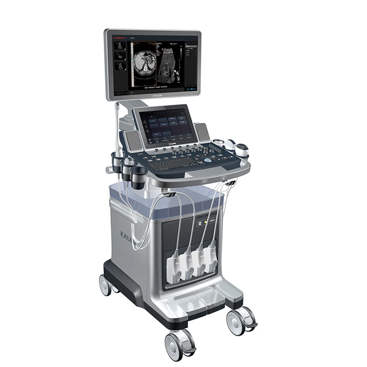
3D/4D дополнительный больница Цветной Допплер ультразвуковой аппарат
- Категория: Ultrasonic, Optical, Electronic Equipment >>>
- Поставщик: Wuhan,J.H.,Bio-Tech,Co.,Ltd.
Поделиться:
Описание и отзывы
Трекер стоимости
| Месяц | Минимальная цена | Макс. стоимость |
|---|---|---|
| Sep-17-2025 | 0.79 $* | 0.81 $* |
| Aug-17-2025 | 0.51 $* | 0.63 $* |
| Jul-17-2025 | 0.8 $* | 0.26 $* |
| Jun-17-2025 | 0.21 $* | 0.47 $* |
| May-17-2025 | 0.42 $* | 0.34 $* |
| Apr-17-2025 | 0.68 $* | 0.43 $* |
| Mar-17-2025 | 0.61 $* | 0.62 $* |
| Feb-17-2025 | 0.60 $* | 0.52 $* |
| Jan-17-2025 | 0.64 $* | 0.63 $* |
Характеристики


4D Optional Hospital Color Doppler Ultrasound machine KAI-X10
Standard configuration(4d) |
1 Full Digital Color Ultrasound Diagnostic System Host , |
2 Display: ≥ 15-inch high-resolution progressive-scan LCD displays , |
3 linear array ultra-high frequency ultrasound probe change (5.0 ~ 12.0MHz) , |
4 convex array broadband probe cavity frequency, the frequency range (4 - 8MHz) ,, |
5 4D probe, |
6 Built-in workstation systems (including software), |
7 ultrasound coupling agent |
8 Power lines |
Options |
multi-frequency convex(heart) probe, multi-frequency convex(rectal)probe, video printer, |
Trolley 4D ultrasound machine Standard configuration:
one mainframe unit of ultrasound;
1pc convex array probe;
1pc high frequency linear array probe;
1pc 4D volume probe
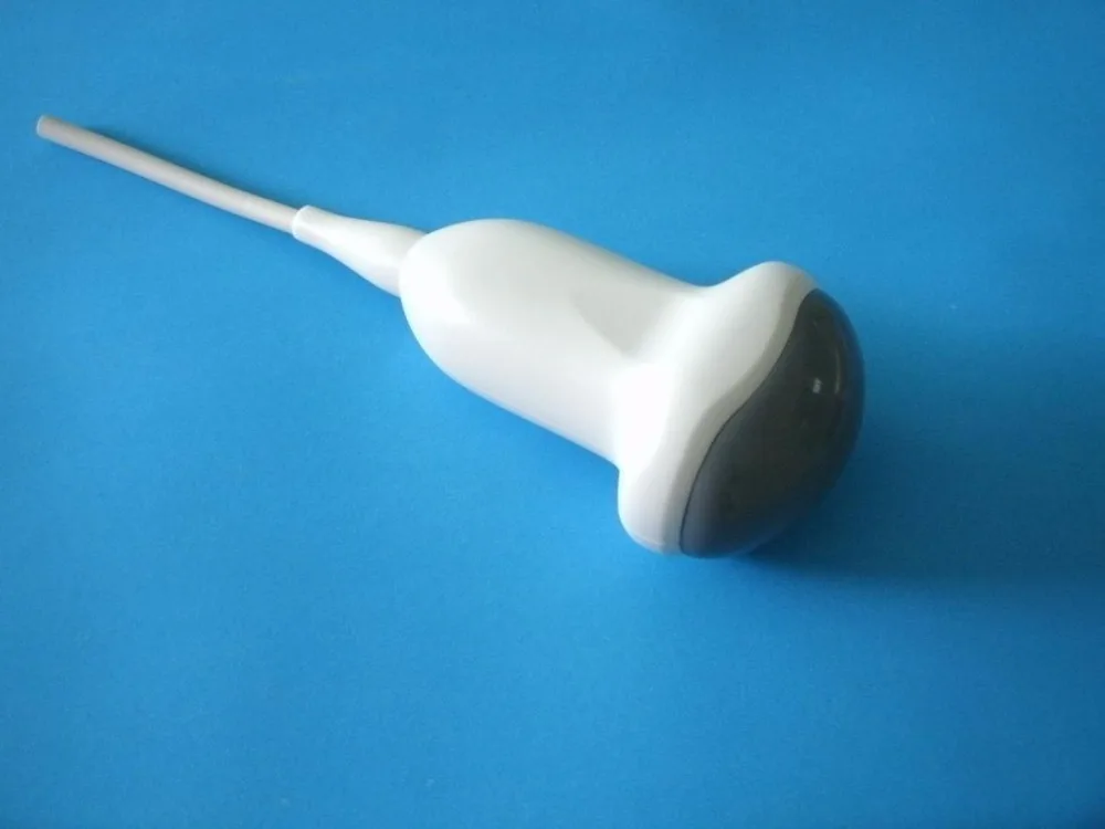
Specification for 4D probe C3.5-128R40C:
center frequency: 3.5MHz
array element: 128
radius of curvature:40.0mm
lateral aperture: 13.0mm
focal length: 80.0±10.0 mm
Product details:
A. Innovative technology ,superior performance
1 Weighted launch technology
The transmission model by the weighted against array element different voltage, pulse, overcome the far-field sound interfere with each other to enhance the trunk, suppress the sidelobe, eliminate artifacts.
2 The dynamic real-time blood flow imaging
Through accurate Doppler echo signal automatic recognition processing, reduce blood vessels and blood flow Doppler signals back to the fold and make within the blood vessels and vascular Doppler blood flow imaging showed more clearly.
3 Pixel focusing technology
Powerful digital beam forming device, is used for controlling pixels focus, improve the accuracy of the spatial resolution, image display fine structure.
4 Ultra wideband beamforming
Ensure the whole piece of information without loss or distortion, effectively improve the image resolution
5 Compound imaging technology
Make the image more smooth nature, more easy to distinguish the smallest details
6 One touch optimization function
Fast image optimization effect, can improve the efficiency of clinical work
7 User-defined parameter Settings
Users can set parameters for clinical diagnosis according to their own needs
8 The application of harmonic technique
The resolution of the images can improve effective, and increase the basis of clinical diagnosis
B. Advanced software, clinical assistant
1 IClear image optimization function
According to actual needs, it can adjust the softness of black/white image, and make the image more clear and reliable performance.
2 Rich and professional report page
Professional obstetric report page contains fetal growth curve comparison table, due date the measurement data, and can add personalized diagnostic template.
3 Built-in workstation
It is equipped with a powerful PC System inside the machine, and the doctor can write a report on the system directly. Furthermore, it can connect various printers.
4 A variety of professional application of the probe
It can connect convex array probe, high frequency probe, cavity probe, probe of heart probe and other professional diagnosis.